Functional neuroimaging of traumatic brain injury with Positron Emission Tomography (PET)
See Levine et al., 2002
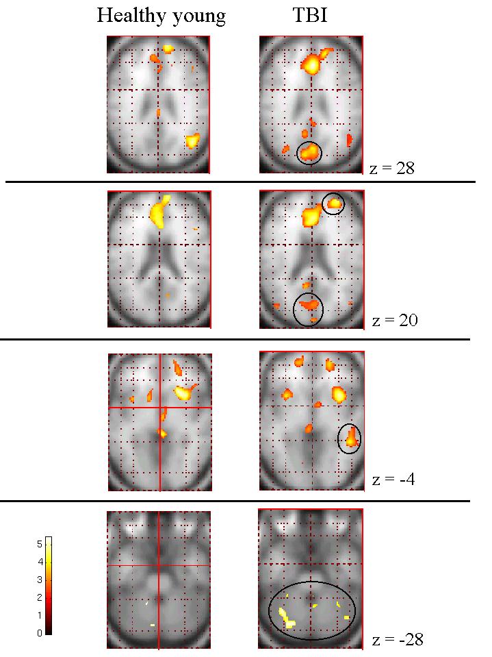
Patients who have made good recovery from significant traumatic brain injury (TBI) show areas of increased brain activity (circled) when performing memory tasks.
These areas of increased activation may represent functional re-organization of brain networks supporting memory as a result of recovery from brain injury. Alternatively, they may represent disinhibition of memory circuits (i.e., less efficent brain activity).
Current studies in our laboratory, led by Danielle Tisserand, Charlene O'Connor, and Craig Easdon are examining the functional neuroanatomy of traumatic brain injury using functional magnetic resonance imaging (fMRI).
See Levine et al., 2002
Patients who have made good recovery from significant traumatic brain injury (TBI) show areas of increased brain activity (circled) when performing memory tasks.
These areas of increased activation may represent functional re-organization of brain networks supporting memory as a result of recovery from brain injury. Alternatively, they may represent disinhibition of memory circuits (i.e., less efficent brain activity).
Current studies in our laboratory, led by Danielle Tisserand, Charlene O'Connor, and Craig Easdon are examining the functional neuroanatomy of traumatic brain injury using functional magnetic resonance imaging (fMRI).


